Hip Arthroscopy

Hip arthroscopy is a surgical procedure that allows doctors to view the hip joint through 2-3 small incisions (cuts) through the skin. Arthroscopy is used to diagnose and treat a wide range of hip problems.

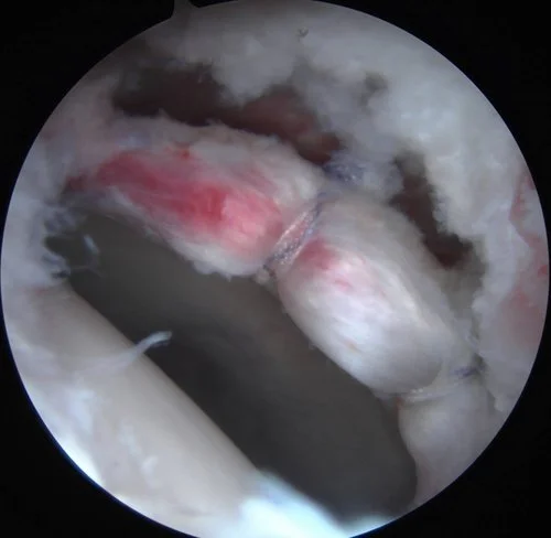
During hip arthroscopy, your surgeon inserts a small camera, called an arthroscope, into your hip joint. The camera displays pictures on a video monitor, and your surgeon uses these images to guide miniature surgical instruments.

Dr. Wang uses the Stryker Pivot Guardian Distraction Table for hip arthroscopy. This is an innovative, post-less table that eliminates groin and nerve complications. For more information on this post-less table, click here.

Hip arthroscopy may relieve painful symptoms of many problems that damage the labrum, articular cartilage, or other soft tissues surrounding the joint. Some of these orthopaedic conditions include:

  • Femoroacetabular impingement (FAI) is a disorder in which extra bone develops along the acetabulum (pincer impingement) or on the femoral head (cam impingement). Most of the time, bone spurs develop on both the acetabulum and femoral head. Over time, repetitive impingement can damage the soft tissues of the hip (labrum and articular cartilage) during movement.

  • Dysplasia is a condition in which the hip socket is abnormally shallow. This puts more stress on the labrum to keep the femoral head within the socket, and makes the labrum more susceptible to tearing.

  • Snapping hip syndromes cause a tendon to rub across the outside of the joint. This type of snapping or popping is often harmless. However, in some cases, however, the tendon is damaged from the repeated rubbing.

  • Synovitis causes the tissues that surround the joint to become inflamed.

  • Loose bodies are fragments of bone or cartilage that become loose and move around within the joint.

  • Hip joint infection